弹性蛋白聚合物(ELP)凝聚层分析的新型高通量方法
New High-Throughput Method for Elastin-like Polymer (ELP) Coacervate Analysis
弹性蛋白聚合物(ELP)凝聚层分析的新型高通量方法
Posted by Michelle Devoe
Michelle Devoe发表
December 2018 — A recent study by researchers from the University of New England and University of New Hampshire has demonstrated that flow imaging microscopy is an accurate, more efficient, and more informative method of elastin-like polymer (ELP) coacervate analysis than standard methods. ELP coacervates are a class of molecules with promising applications in drug delivery vehicles, tissue engineering, environmental remediation, and more. ELP coacervate architecture is stimuli-responsive and highly tunable, making them ideal for the above-mentioned applications.
2018年12月 - 新英格兰大学和新罕布什尔大学的研究人员Z近的一项研究表明,与标准方法相比,流动成像显微镜是一种准确,更有效,信息更丰富的弹性蛋白样聚合物(ELP)凝聚分析方法。 ELP凝聚层在作为药物递送的载体,组织工程,环境修复等方面具有广阔的应用前景。 ELP凝聚层结构具有刺激响应性和高度可调性,使其成为上述应用的理想选择。
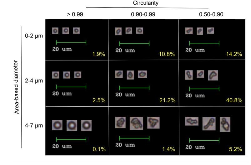
ELP coacervates imaged by the FlowCam. Size and circularity, two of the 40+ properties that can be measured by the FlowCam, is used to sort coacervates. Image from Marvin et al. (2018).
ELP凝聚了由FlowCam成像的图像。 尺寸和圆度是FlowCam可以测量的40多个属性中的两个,用于对凝聚层进行分类。 图片来自Marvin等。(2018)。
Standard methods for ELP coacervate analysis are indirect and cumbersome. Data from Visible-UV spectrophotometer turbidity measurements and dynamic light scattering (DLS) size measurements are superimposed to evaluate the formation of micron-scale aggregates, and particle geometry is constructed indirectly from diffusion data. Imaging analysis methods such as optical, electron, and scanning probe microscopy are useful methods to acquire size and shape data, however it is time-consuming and laborious to acquire a large enough sample size to get statistically significant data.
ELP凝聚层分析的标准方法是间接和麻烦的。 来自可见 - 紫外分光光度计浊度测量和动态光散射(DLS)尺寸测量的数据被叠加以评估微米级聚集体的形成,并且颗粒几何形状间接地由扩散数据构建。 诸如光学,电子和扫描探针显微镜之类的成像分析方法是获取尺寸和形状数据的有用方法,然而获得足够大的样本大小以获得统计上有效的数据是耗时且费力的。
In this study by the University of New England and University of New Hampshire, the size, morphology, and behavior of ELP coacervates subjected to various solvent conditions were measured and observed using the FlowCam particle analyzer. Results were validated by comparison with DLS and atomic force microscopy analyses. Flow imaging microscopy was demonstrated to be a successful method for ELP coacervate analysis. Additionally, flow imaging microscopy reported additional findings that were not measured using the DLS, microscopy, or Visible-UV spectrophotometry.
在新英格兰大学和新罕布什尔大学的这项研究中,使用FlowCam颗粒分析仪测量和观察经受各种溶剂条件的ELP凝聚层的尺寸,形态和行为。 通过与DLS和原子力显微镜分析的比较验证结果。 流动成像显微镜被证明是ELP凝聚层分析的成功方法。 此外,流动成像显微镜提供了DLS,显微镜或可见紫外分光光度法不能获得的结果。
相关产品
全部评论(0条)
推荐阅读
-
- 高通量毛细管电泳技术,开启蛋白药物分析新纪元
- 高通量毛细管电泳技术,开启蛋白药物分析新纪元
-
- Copure | 蛋白沉淀法如何实现高效率、高通量处理样本?
- 专为去除生物液体样品(血浆、血清或血液)中的蛋白质而设计!
-
- 快看!这才是聚合物快速分析的正确打开方式
- 快看!这才是聚合物快速分析的正确打开方式
-
- 热裂解APGC QTof在聚合物分析中的应用
- 热裂解APGC QTof在聚合物分析中的应用
-
- 热裂解APGC QTof在聚合物分析中的应用
- 热裂解APGC QTof在聚合物分析中的应用


①本文由仪器网入驻的作者或注册的会员撰写并发布,观点仅代表作者本人,不代表仪器网立场。若内容侵犯到您的合法权益,请及时告诉,我们立即通知作者,并马上删除。
②凡本网注明"来源:仪器网"的所有作品,版权均属于仪器网,转载时须经本网同意,并请注明仪器网(www.yiqi.com)。
③本网转载并注明来源的作品,目的在于传递更多信息,并不代表本网赞同其观点或证实其内容的真实性,不承担此类作品侵权行为的直接责任及连带责任。其他媒体、网站或个人从本网转载时,必须保留本网注明的作品来源,并自负版权等法律责任。
④若本站内容侵犯到您的合法权益,请及时告诉,我们马上修改或删除。邮箱:hezou_yiqi


参与评论
登录后参与评论